Dr. Edward Gerstenfeld, Professor of Medicine and Chief of Cardiac Electrophysiology at UCSF, and the BEAT-AFib Study team have been recruiting patients with a high burden of PACs to look into how PACs can lead to the development of AFib. But, what are PACs exactly?
Premature atrial contractions (PACs) are a common arrhythmia that many people are likely to experience in their life. PACs can be explained as extra beats in the top chambers of the heart, called the atria. They can sometimes appear when we are stressed or when we drink caffeine or alcohol, for example, but the heart generally returns to its normal rhythm quickly. The extra beat occurs when the heart’s electrical rhythm is generated from somewhere within the walls of the atria instead of using the heart’s own natural pacemaker, the sinoatrial (SA) node.1 PACs can feel like a stronger beat or feel like your heart skipped a beat; otherwise, they do not cause much discomfort, some people do not experience any symptoms.
In contrast, atrial fibrillation (AFib) is when the atria quiver due to the heart’s rhythm taking multiple paths instead of sending one clear signal. AFib can be found when scarring in the heart creates obstacles for the electrical impulse that it now has to navigate through. This has been described as feeling like a flutter or a pounding in the chest.
Although PACs are considered harmless and normal in most people, it is when PACs occur at a higher rate where they have the ability to cause electrical and structural changes to the heart.2 The heart’s rhythm can begin to slow and the atria can enlarge and develop scarring. When the heart’s rhythm is slowed down and the heart muscle develops scarring, it makes it more difficult for the rhythm to take the appropriate time and route through the heart. These electrical and structural changes caused by PACs have been shown to create an environment ideal for causing AFib.
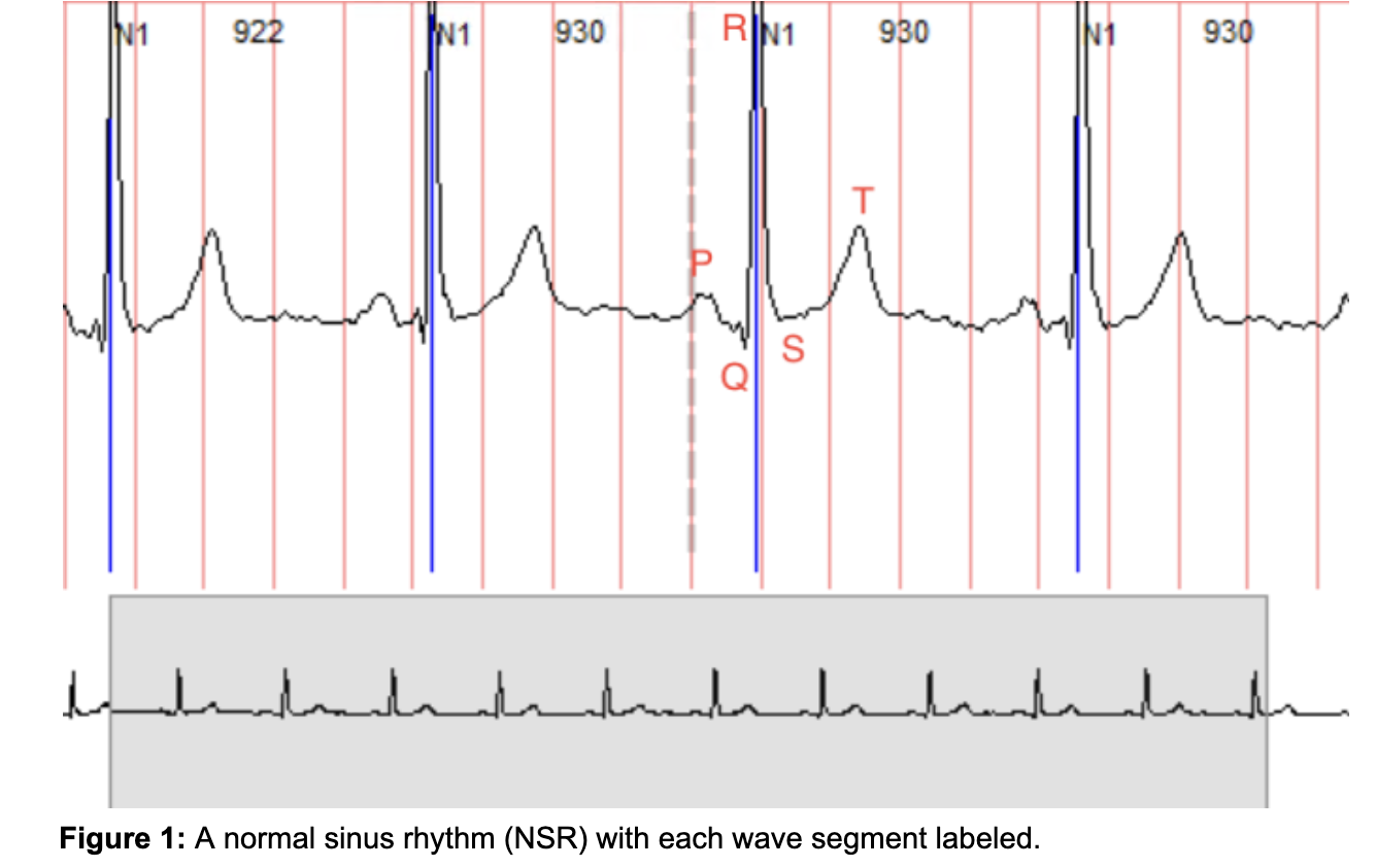
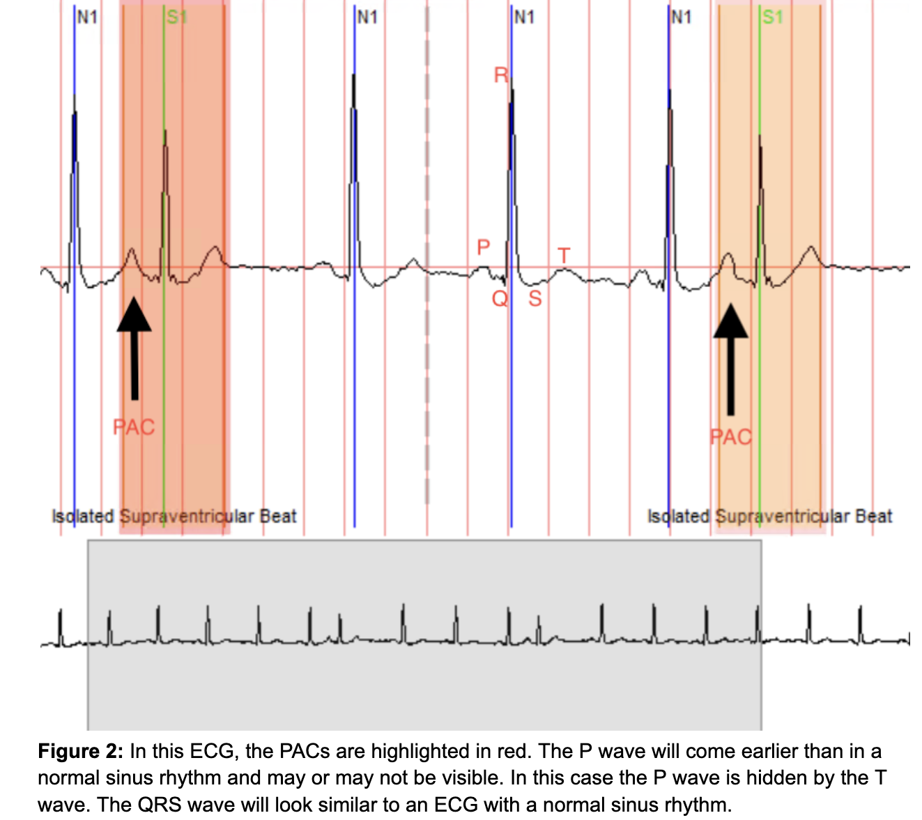
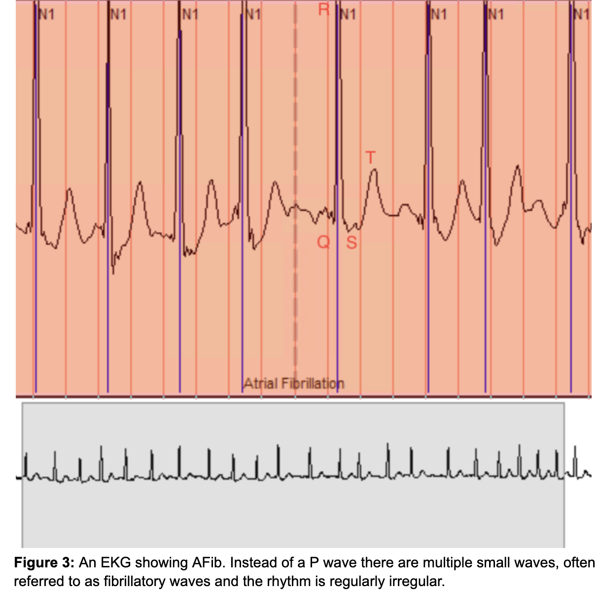
An ECG can show us the changes PACs and AFib cause to the heart’s rhythm. Our previous article about ECGs summarizes the components of an ECG tracing: “an ECG consists of 3 main waves that are examined: a P wave, which represents the electrical activity (depolarization) of the atria, a QRS wave that represents the ventricular (bottom chambers) electrical activity, which is much larger than the atria and creates a much larger wave, and the T wave which shows the return of electrical activity in the ventricles to baseline (repolarization).”3 Figure 1 (below) shows an ECG tracing of the heart’s normal rhythm. Figure 2 shows an ECG tracing with PACs (highlighted in the red areas). When comparing the two figures, you will note that the PACs occur earlier in the cardiac cycle than in Figure 1. Figure 3 shows an ECG tracing containing AFib. In AFib, the P wave is not present and instead is replaced by small rapid ‘fibrillating’ waves and the rhythm is regularly irregular because the heart’s ventricles are not completely contracting and relaxing. This leads to symptoms of palpitations, lightheadedness, etc.
It is with your continued participation and contribution that we will all further our knowledge about PACs and their relationship with AFib. Through the collection of biomarkers, we will better understand how and why PACs are occurring and the changes accompanying them that lead to the development of AFib.
Read some of our UCSF collaborators’ papers about new findings about the relationship between PACs and AFib here.
References
- Heaton, J., & Yandrapalli, S. (2021). Premature Atrial Contractions. PubMed; StatPearls Publishing. https://www.ncbi.nlm.nih.gov/books/NBK559204/
- Higuchi, S., Voskoboinik, A., Im, S. I., Lee, A. C. H., Bahorik, A. L., Arbil, A., Afzal, J., Marcus, G. M., Stillson, C., Bibby, D., Abraham, T. P., Wilson, E., Gerstenfeld, E. P., & Olgin, J. (2023). Frequent Premature Atrial Contractions Lead to Adverse Atrial Remodeling and Atrial Fibrillation in a Swine Model. Circulation. https://doi.org/10.1161/circulationaha.123.065874
- Namjou Khaless, A., (2023). The 15-minute High Resolution Signal Average ECG. Eureka; BEAT-AFib Study. Retrieved December 7, 2023, from https://info.eurekaplatform.org/15-min-ecg/