What is an Echocardiogram?
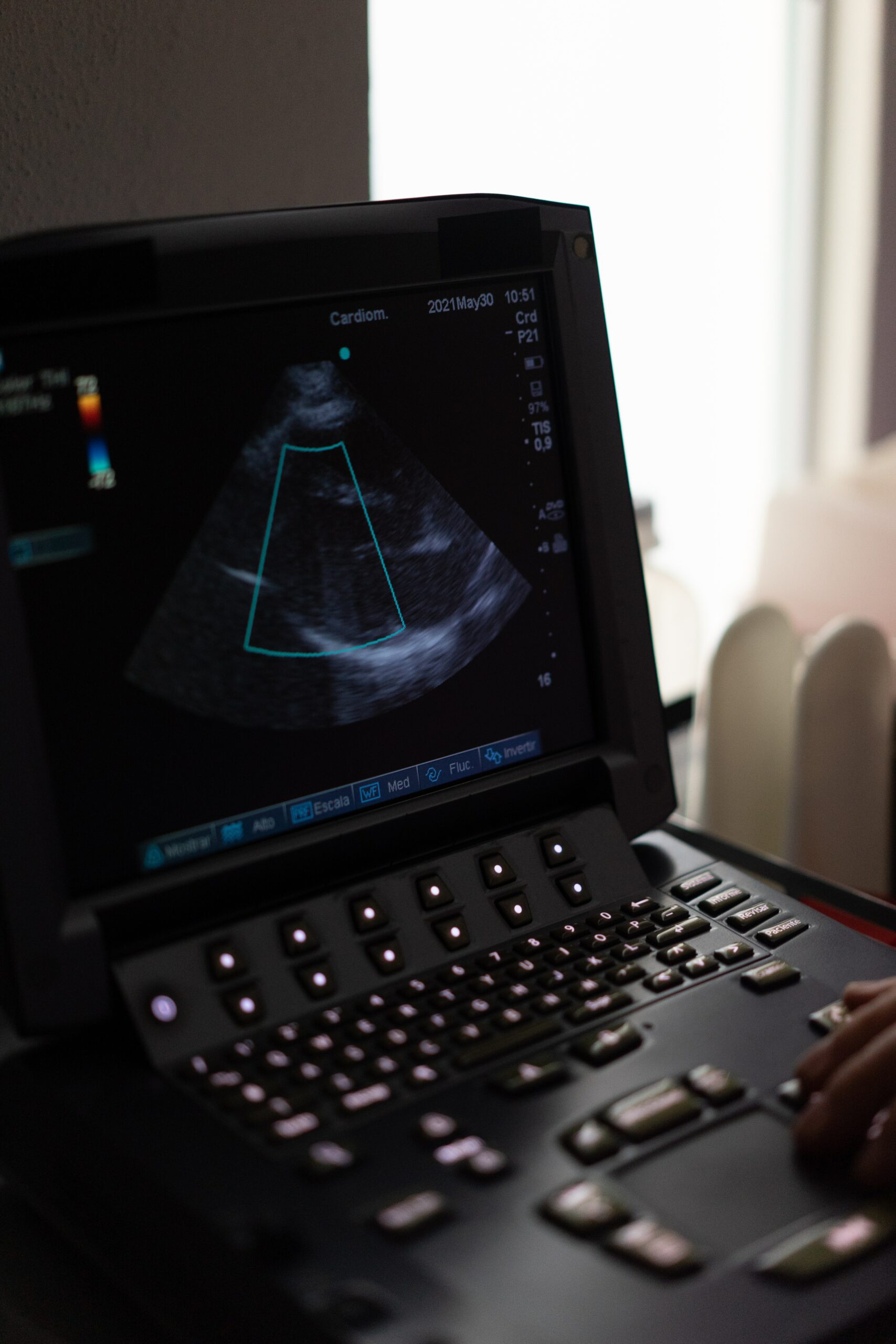
As part of the BEAT-AFib Study, participants undergo an echocardiogram (also referred to as a cardiac echo, an echo, or an ultrasound of the heart). This common test allows us to visualize the heart’s structure and the flow of blood through the heart’s chambers and valves.
What are we looking for?
We are interested in a variety of different features of the heart and how they are functioning. We are able to obtain so much information from the echo, so we will use the left atrium (LA), one of the upper chambers of the heart, as an example. During the echo, we not only take pictures of the atrium from various angles, but we can also see how the LA is functioning; i.e. we can see, in real-time, the LA filling up with blood and then contracting and pushing it to the left ventricle. We are then able to calculate how well the LA is working.
There is increasing evidence that any strain on the functioning of the LA is associated with the development and progression of atrial fibrillation (AFib), and so we are hoping to collect LA strain data on all of our patients and compare the measurements from our AFib patients with our non-AFib patients. Beyond the LA, we take pictures and measurements of the other three chambers of the heart as well. The measurements and calculations performed with the LA is just one example of how ultrasound imaging can be used to further our understanding of AFib.